First image shows cell structure of garlic peel taken from surface of a clove. The next pic compares it with cell structure of a peel taken from the interior. We can see cells are elongated in the interior section.
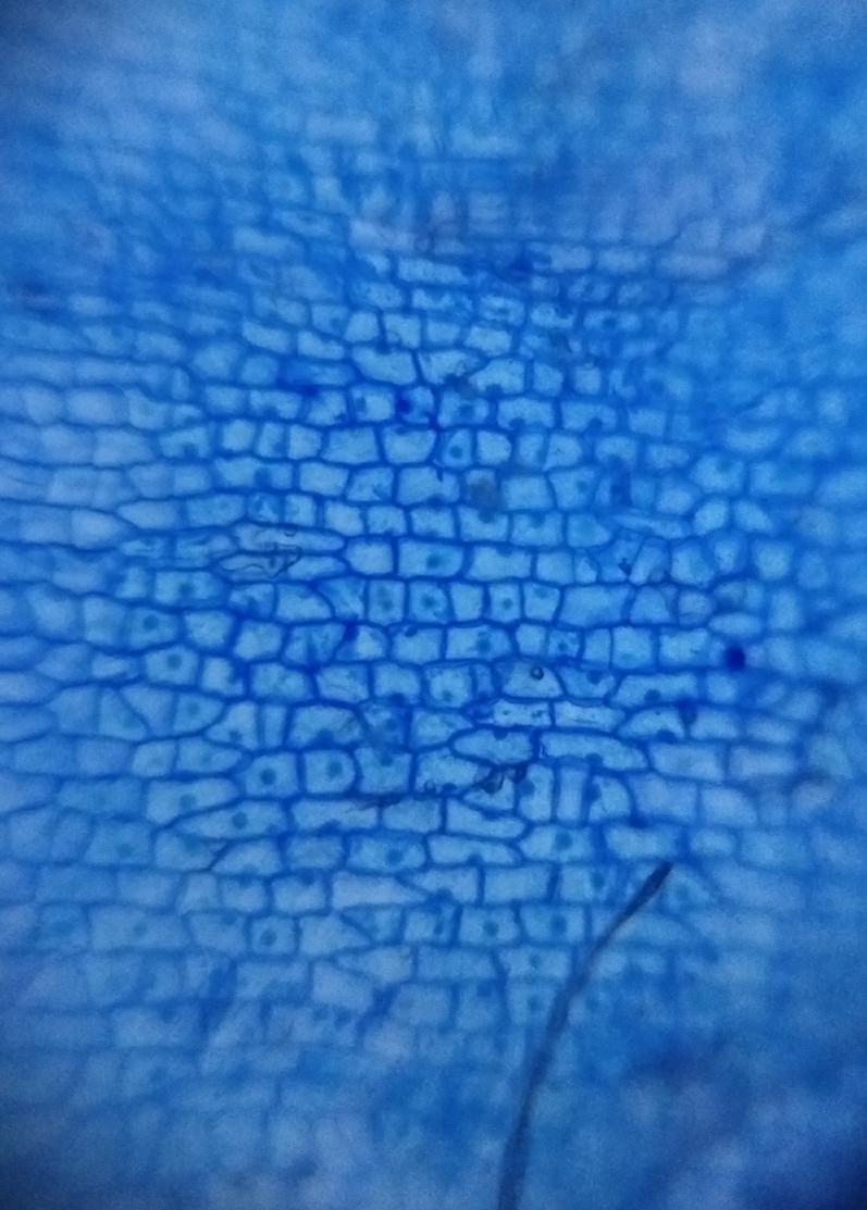
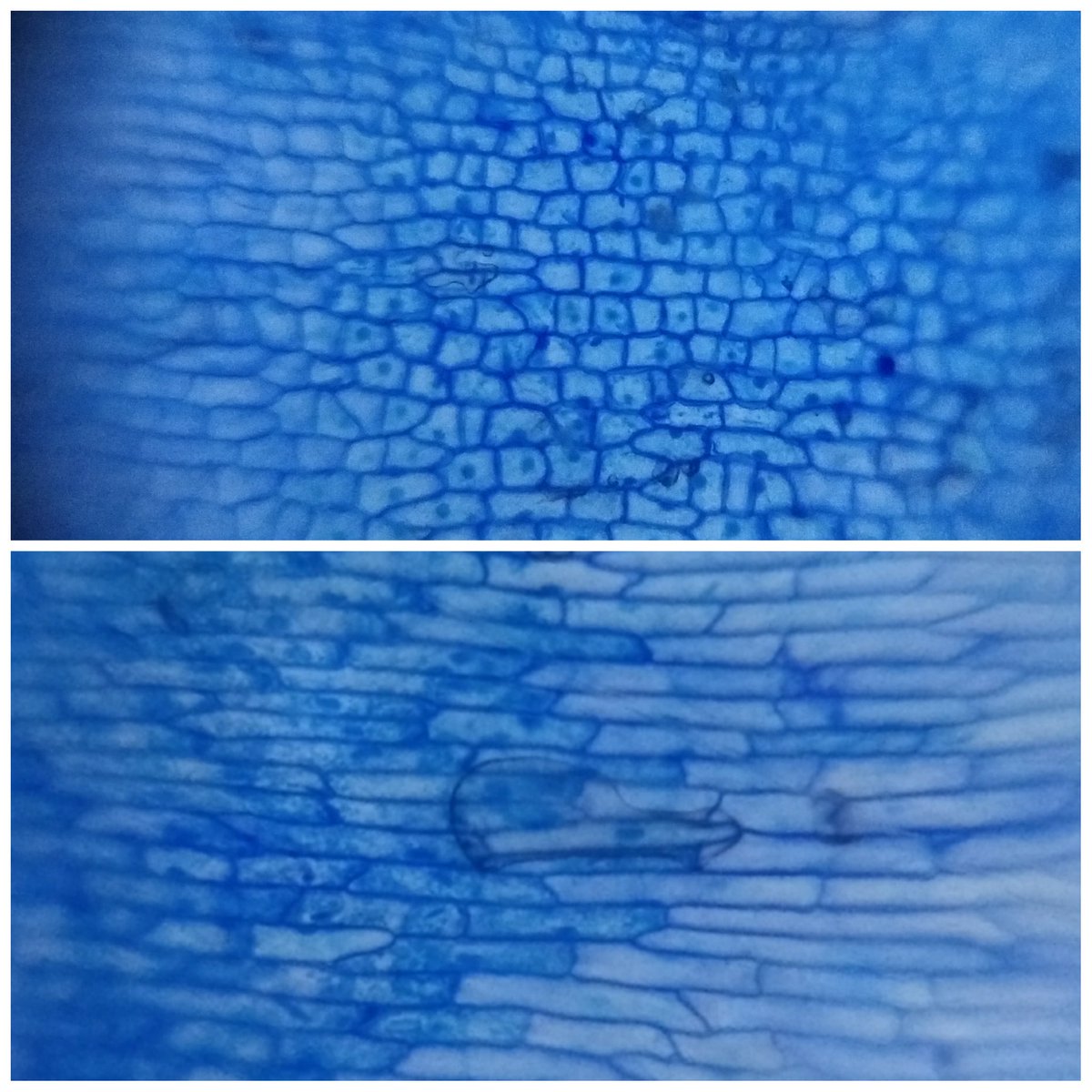
First image shows cell structure of garlic peel taken from surface of a clove. The next pic compares it with cell structure of a peel taken from the interior. We can see cells are elongated in the interior section.


Another pic of garlic peel taken from surface of a garlic clove using blue stain (methelyene blue) obtained from aquarium store. Toward the bottom of the pic we see some cells filled with cytoplasm and nuclei.
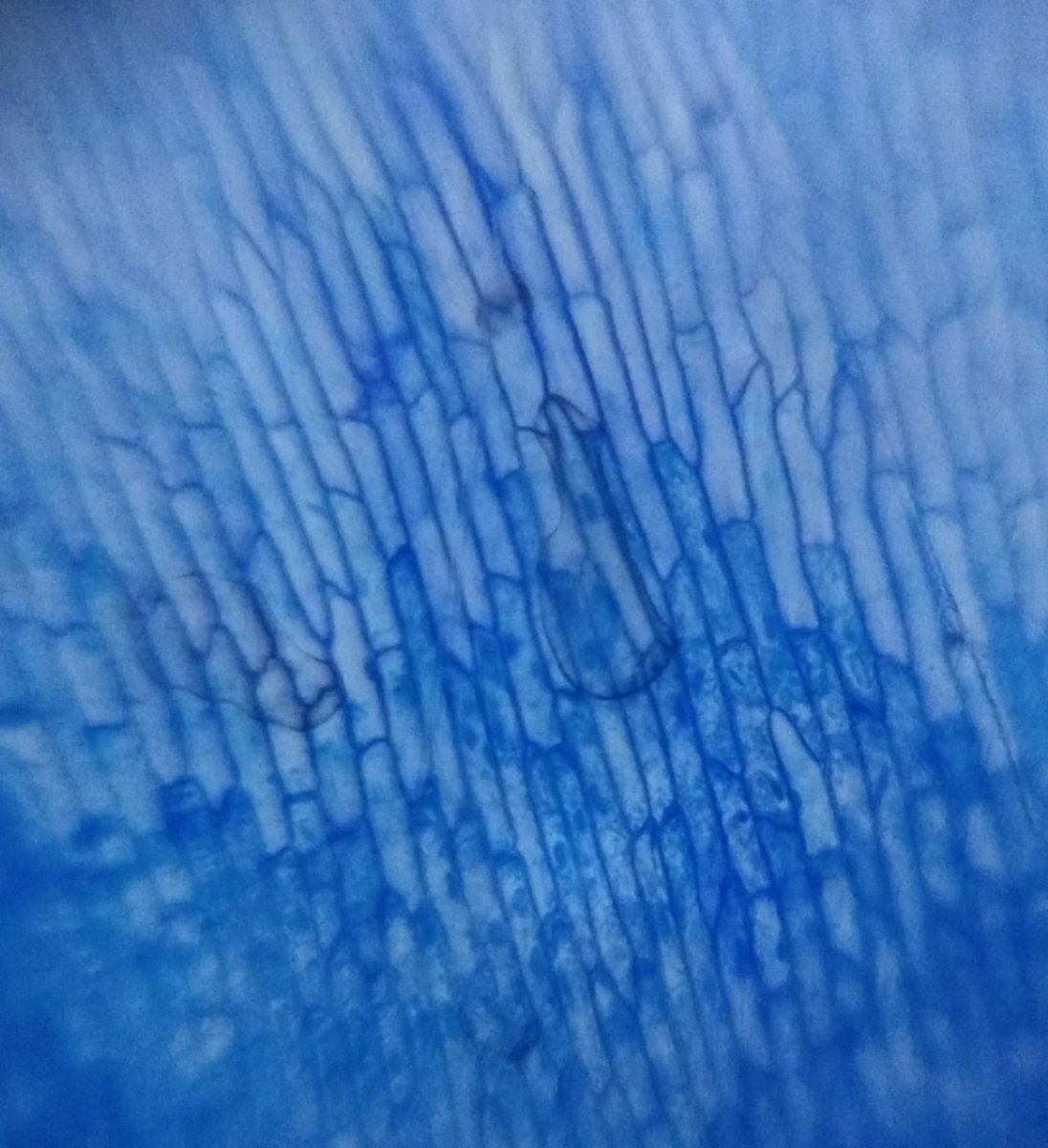
Images of peel taken frm center part of garlic clove . Methelyene Blue used to stain was bought from local aquarium store (the bottle it came in doesn’t specify the chemical name).The pic shows empty cells bcause I think they were damaged during peeling.
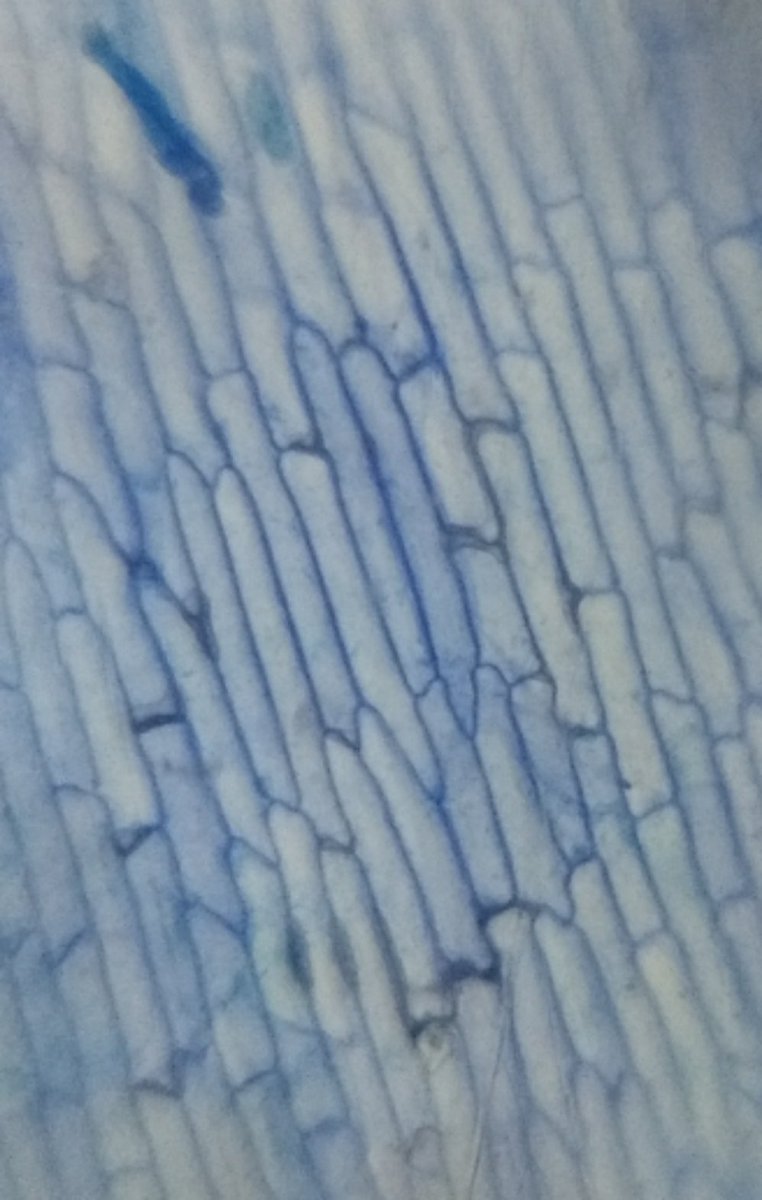
Cell structure of garlic clove. I used antiseptic iodine solution available in medical stores for staining.
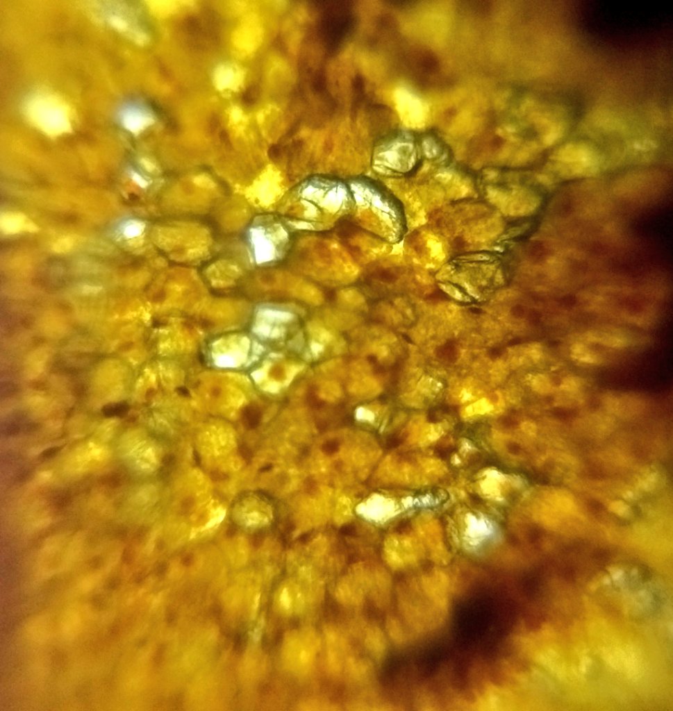
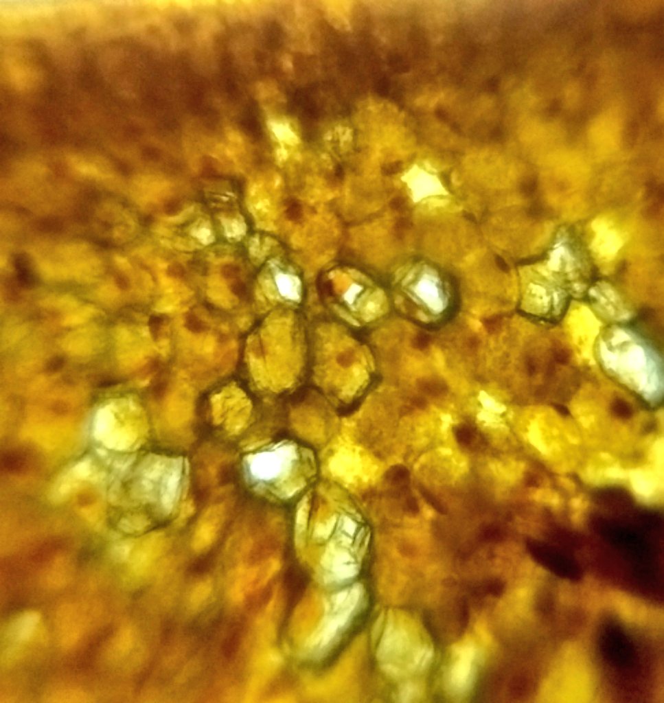
Hibiscus pollen grains seen through a foldscope.
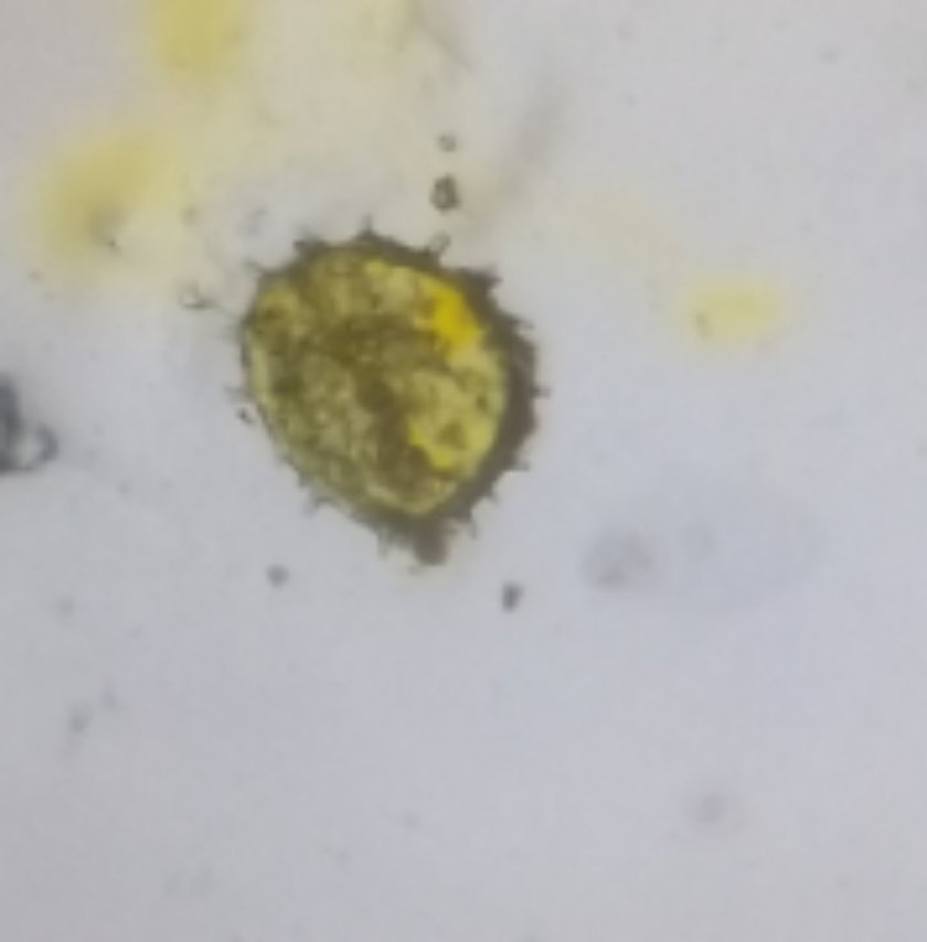
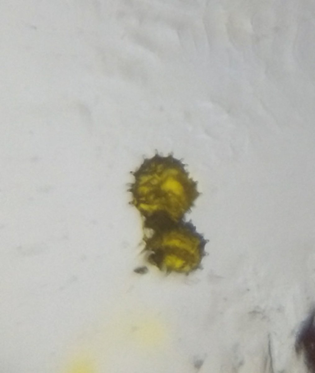
Observations of a cliantro flower using a foldscope. The first image I ‘think’ is that of ag anther. I was not able to identify the part in second image, but it looks cool 😁
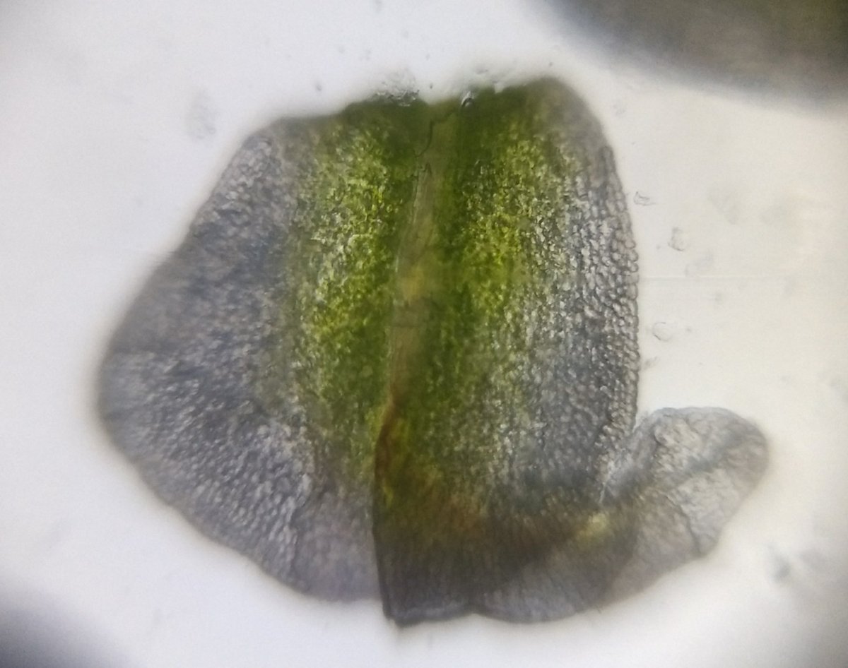
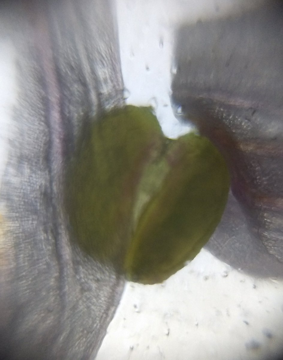
Looks like I was finally able to see stomata using Foldscope. This was epidermal peel of onion leaf.
Any suggestions for easy to obtain staining agents in India. I feel staining might have made a big difference !
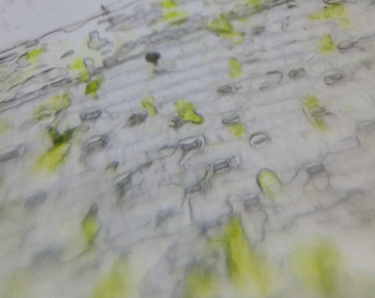
Attempts at observing plan stem of cilantro plant using a Foldscope . I feel something is lacking. Either I am getting cross sections wrong or need to use proper staining agent. Here used red food color, but its not helping it seems.
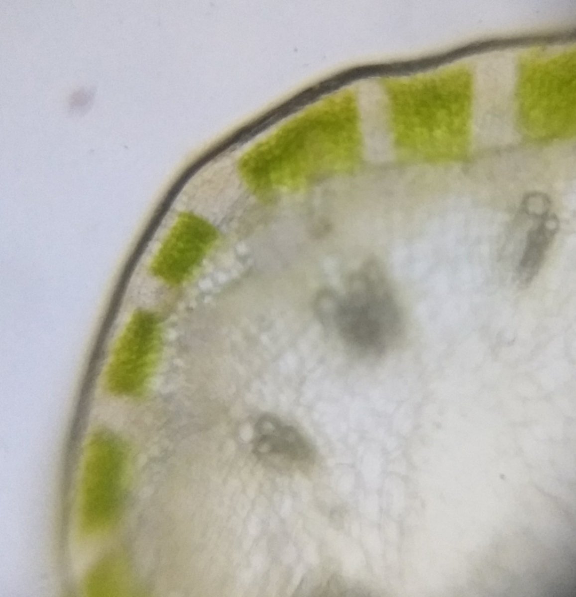
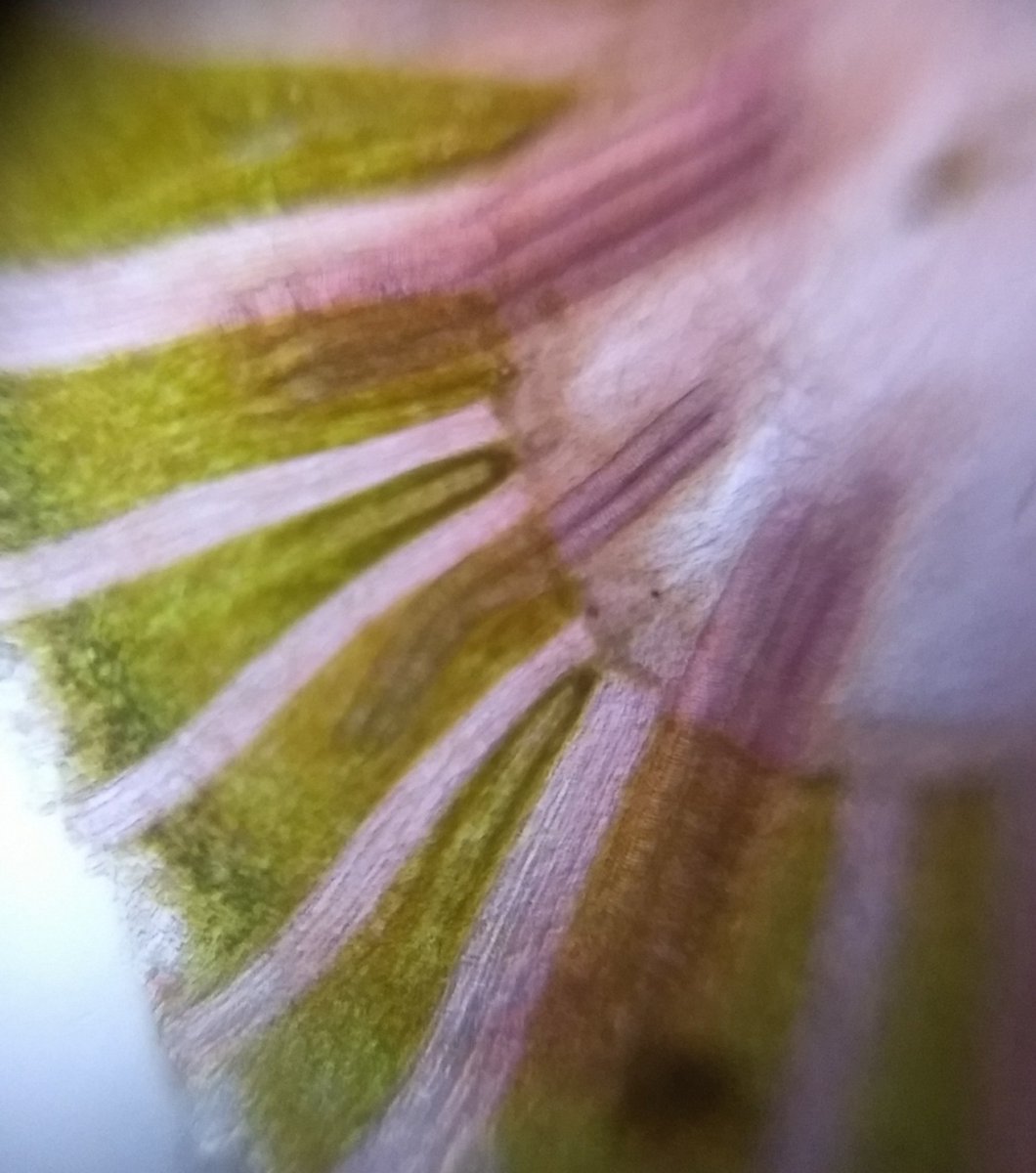
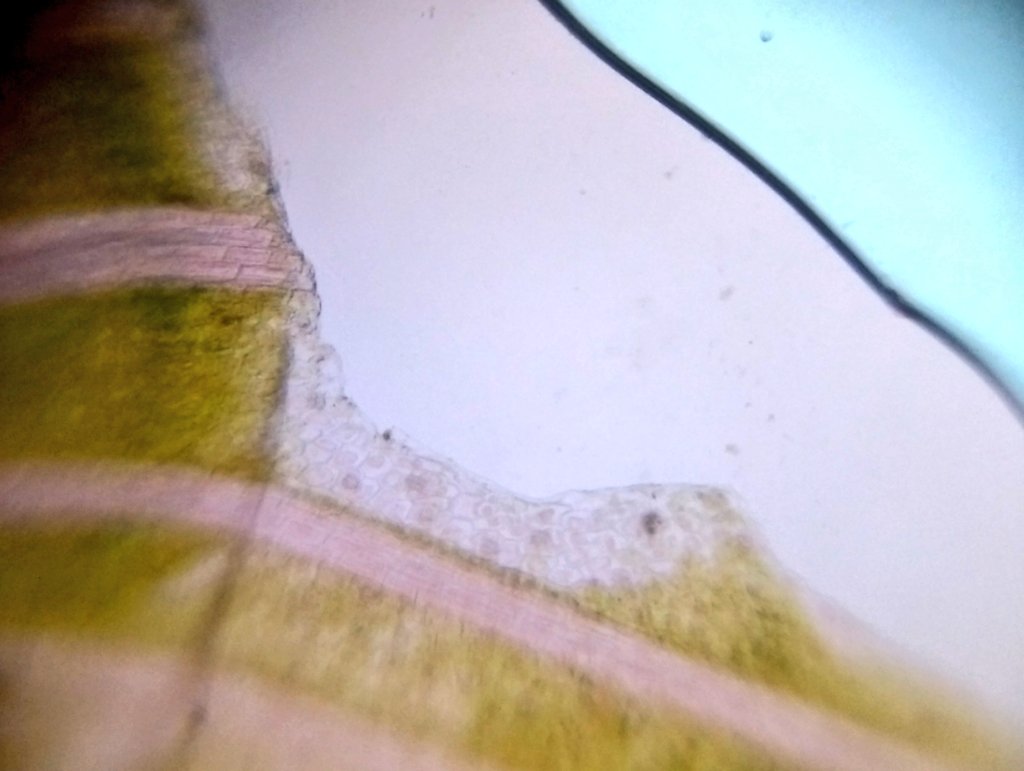
Pollen grains from a Spider Lily. Amazed at the details I could see. Oval structures with reticulate surface can be seen.
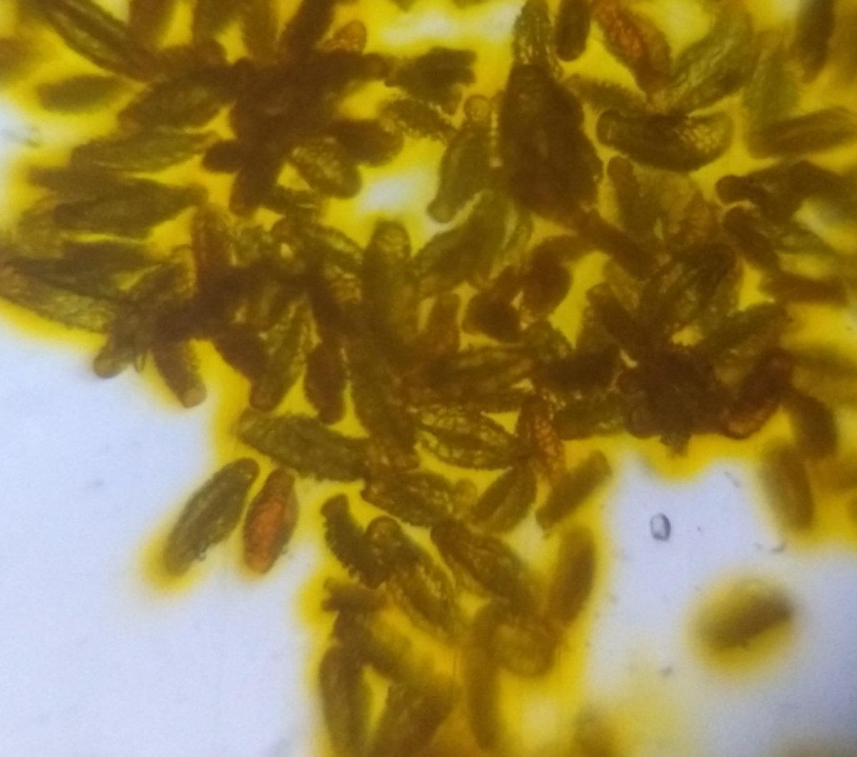
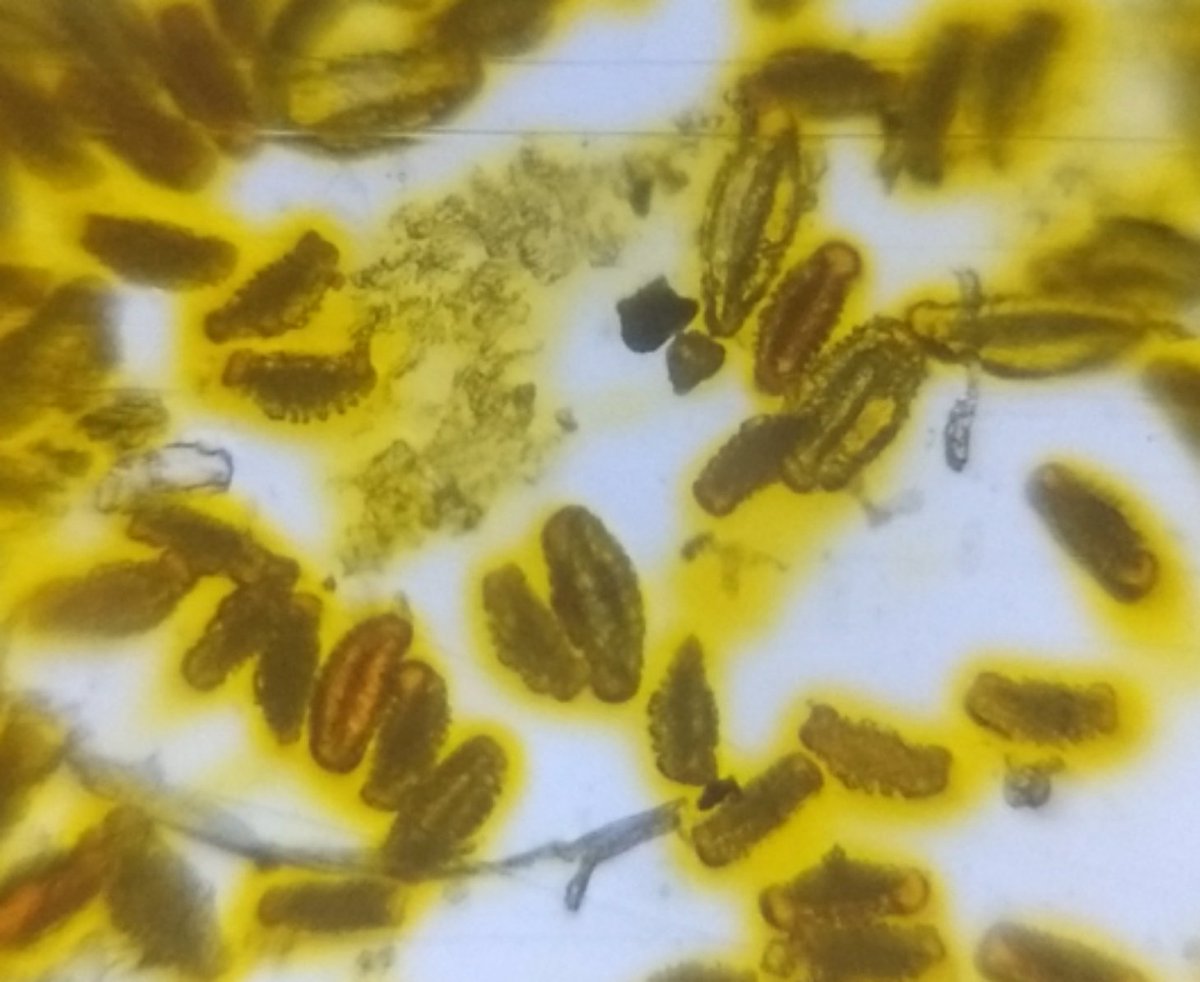
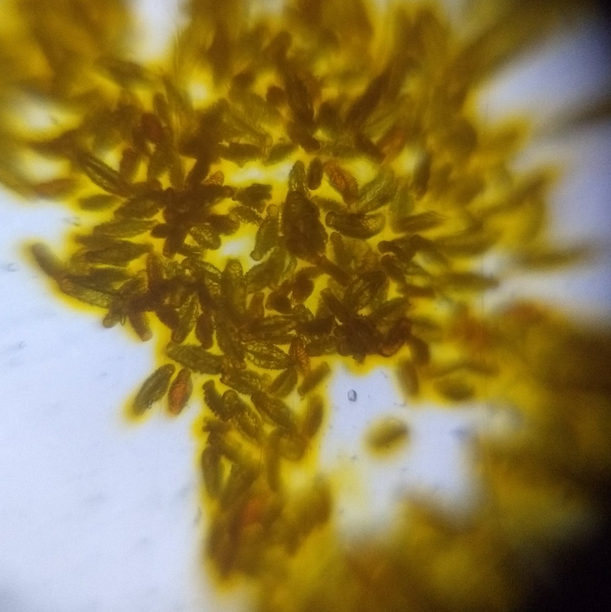
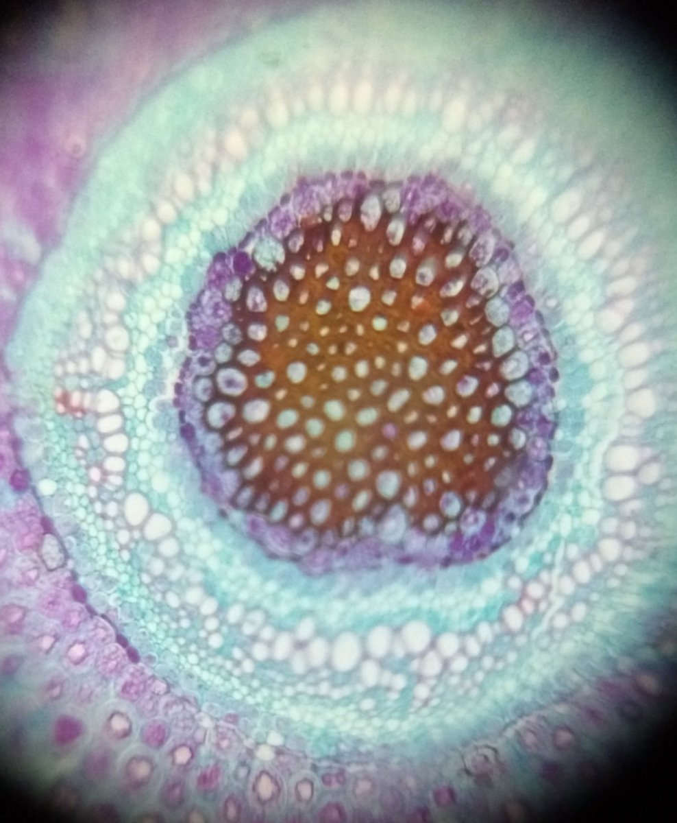
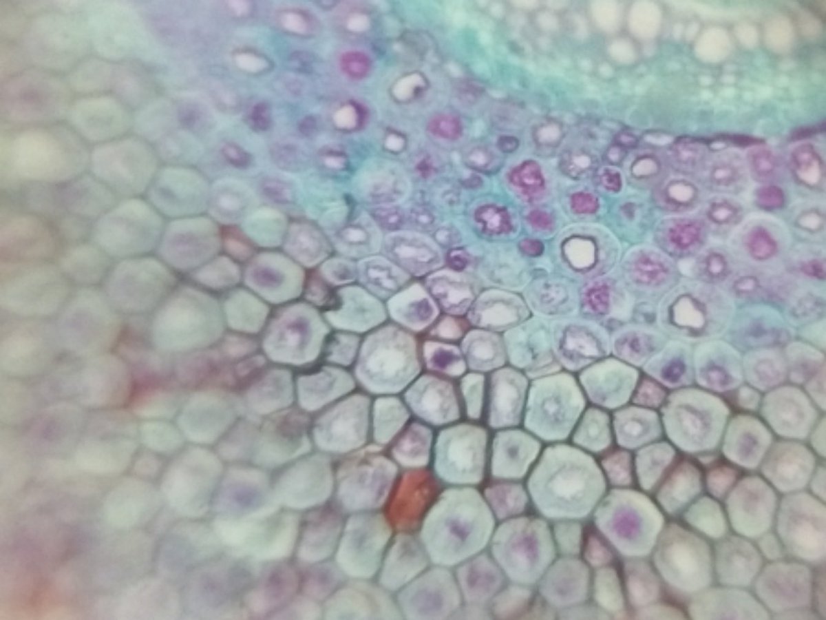
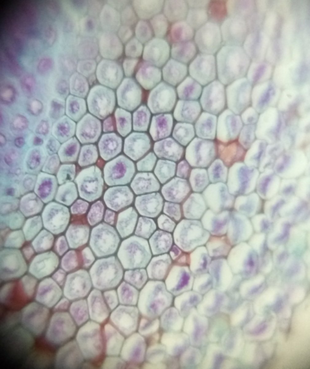
Images of a fern rizome (from a prepared slide that I purchased along with Foldscope)
