Created some artwork using images taken by me using my Foldscope and a technique called Neural Style Transfer ( first image uses Rizhoid cell image and the second one uses garlic cell images, both taken using a Foldscope !
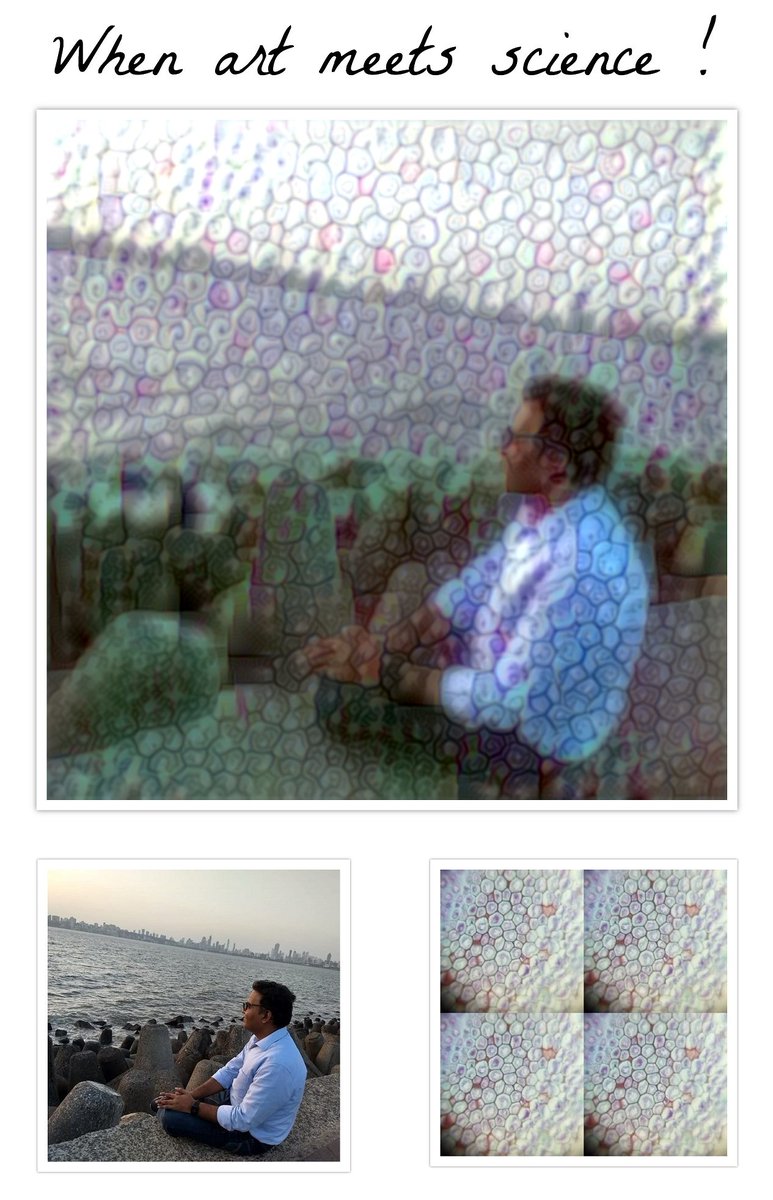
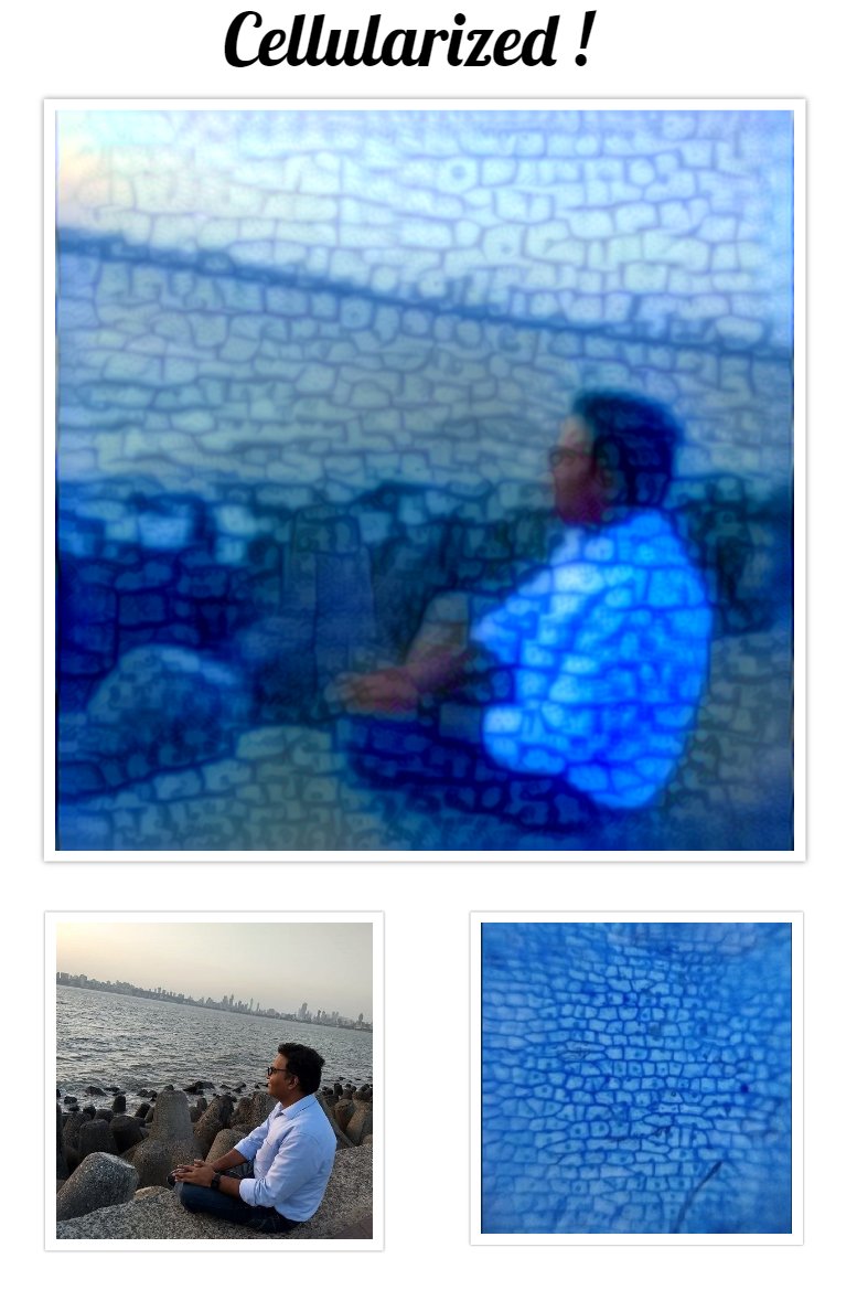
Created some artwork using images taken by me using my Foldscope and a technique called Neural Style Transfer ( first image uses Rizhoid cell image and the second one uses garlic cell images, both taken using a Foldscope !


Found this guy when observing soil sample using Foldscope . Doesn’t look like a colpoda (I could be wrong). I find its internals very interesting. Had a tough time capturing video because this guy was really fast !
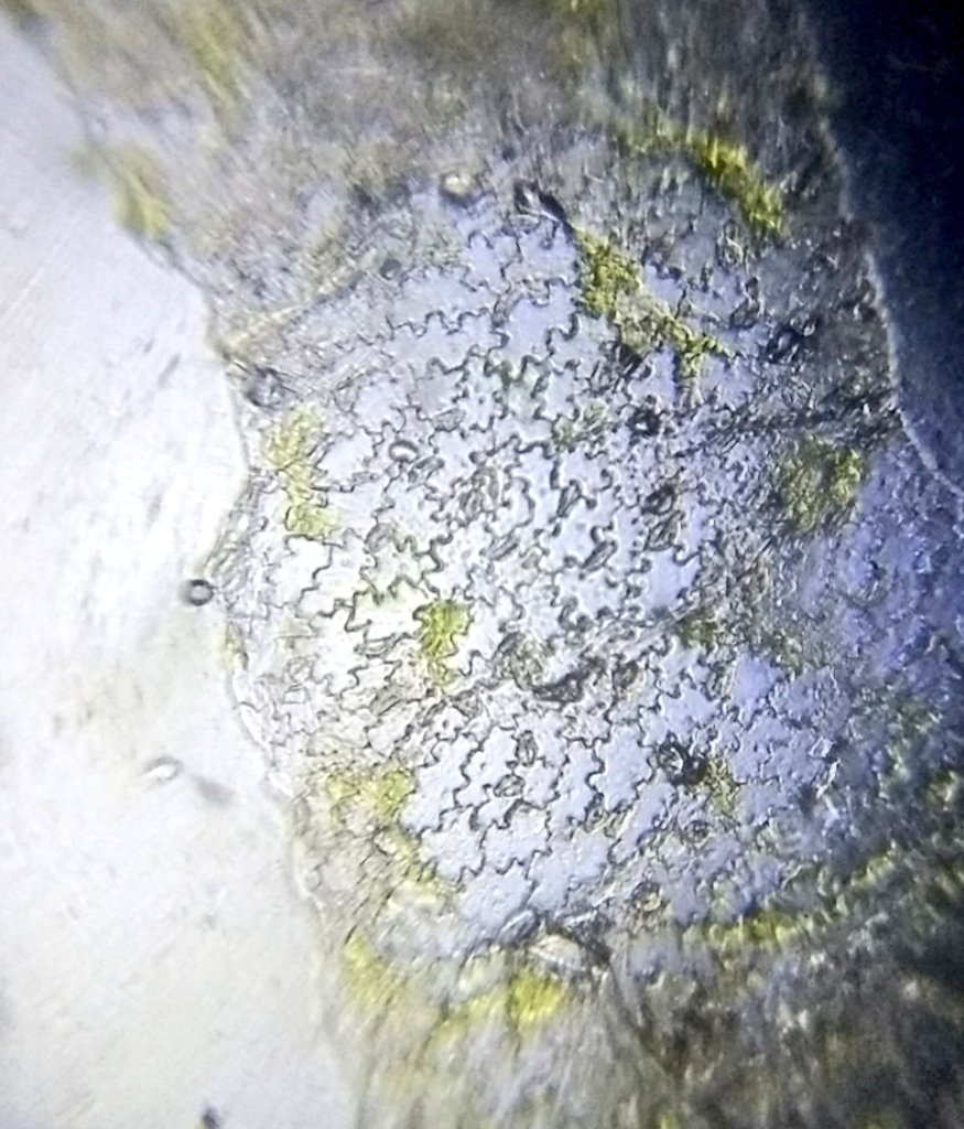
Cells of Fenugreek (Methi) leaf seen using a foldscope. I feel stomata are clearly visible in the first image.
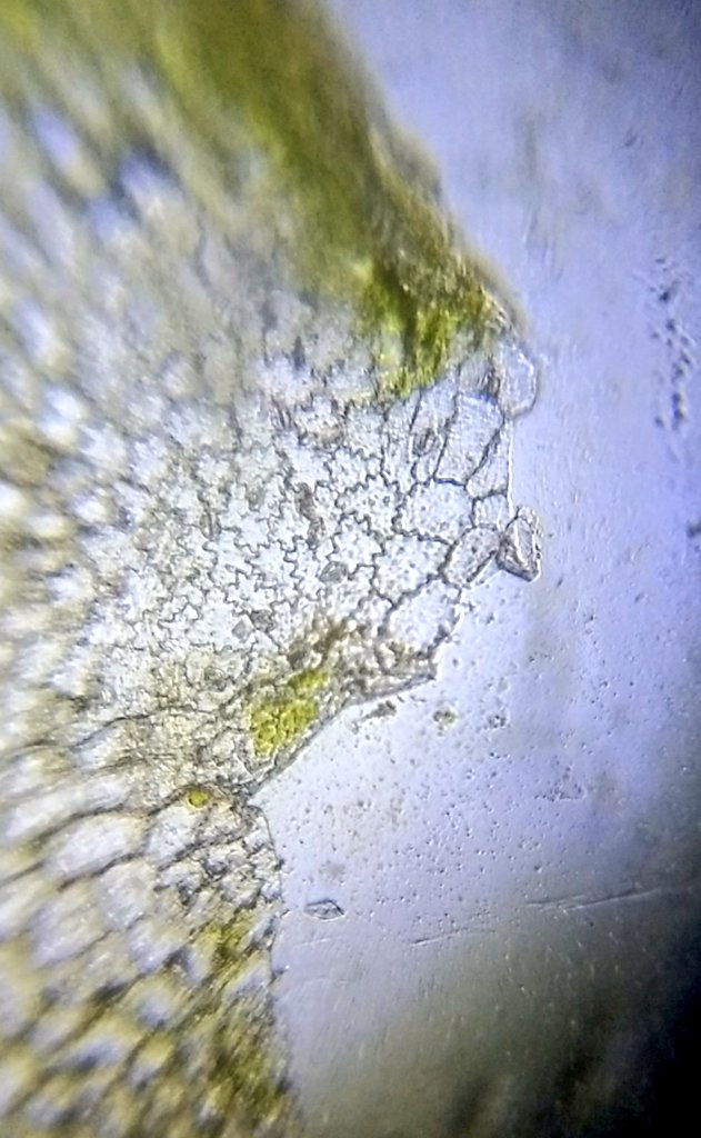
Tiny glass bead like structures on a plant seen through a Foldscope . Looks like these are some kind of eggs.
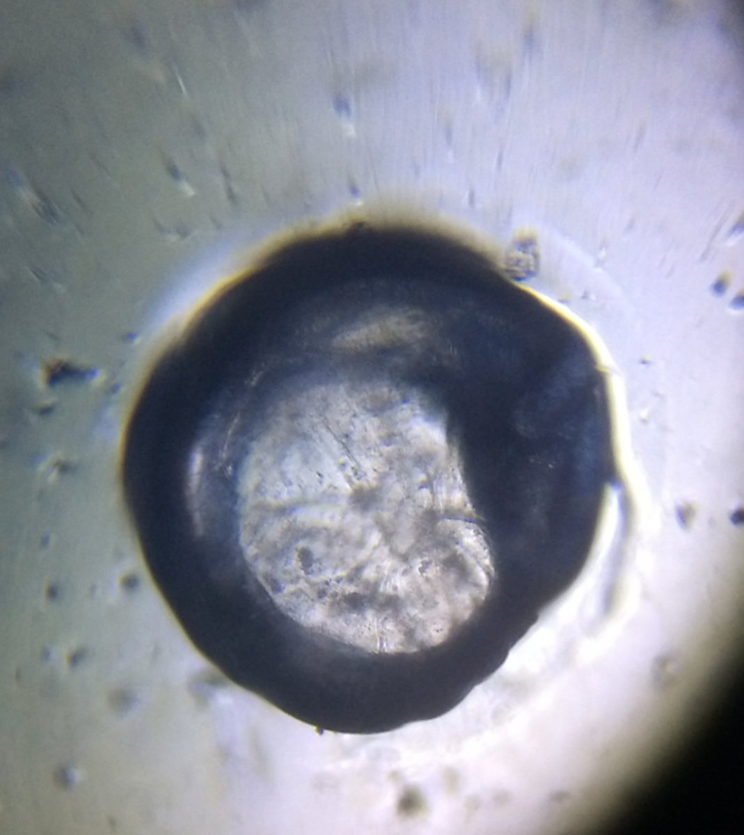
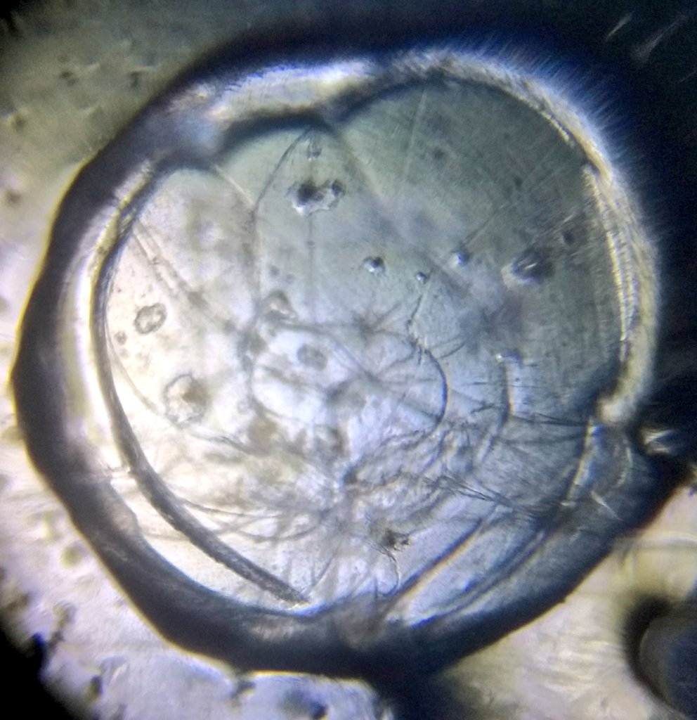
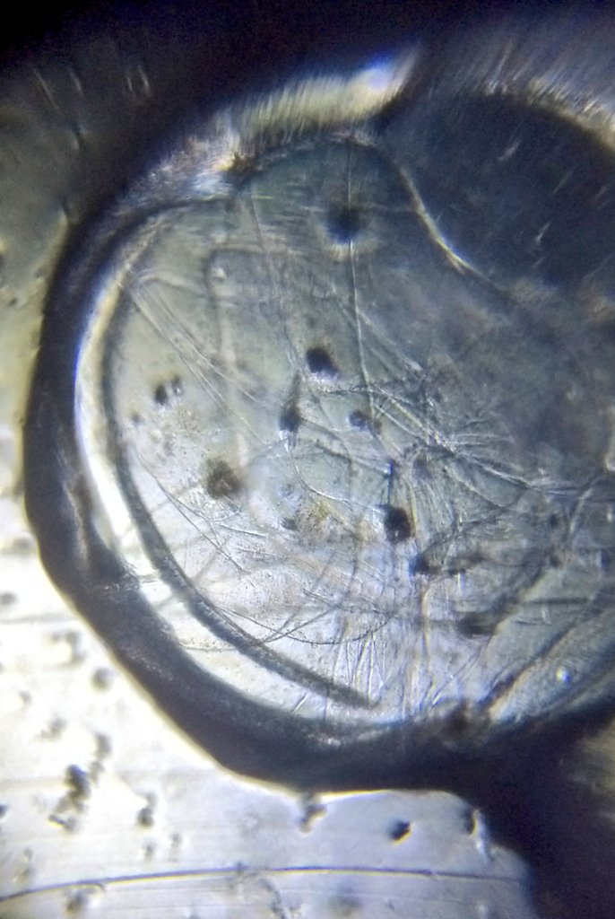
Another video of the amoeba seen via Foldscope. Foldscope is simply awesome. Watch carefully, you should see two jelly like structures towards center left of screen moving downwards.
Had a last look before destroying a sample and look what I found ! Looks like couple of amoebas having fun with ciliates.
See closely and you should be able to see cilia on the round shaped organism. The best way to picture these using Foldscope in my opinion is to trap them in debris of vegetation and use digital zoom.
More fun with Foldscope and ciliates
Something interesting in water sample observed using Foldscope.
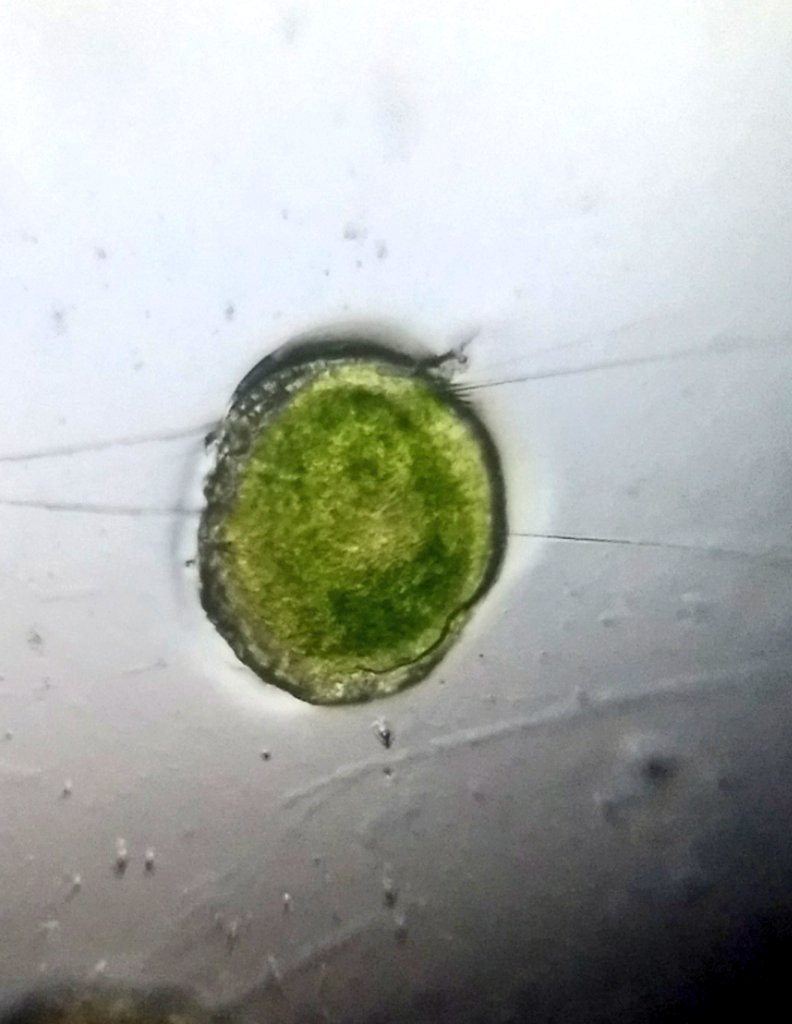
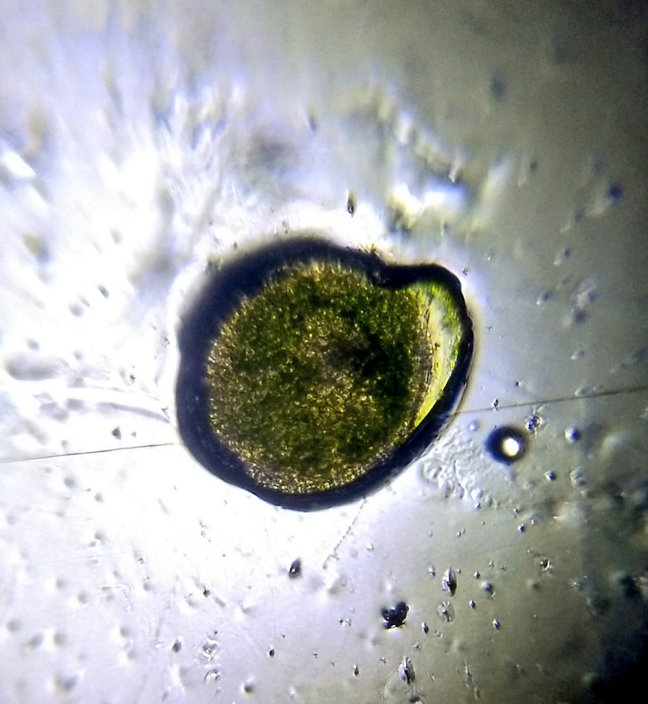
This was not moving and I did come across few more identical structures in the sample. This was biggest of them all.
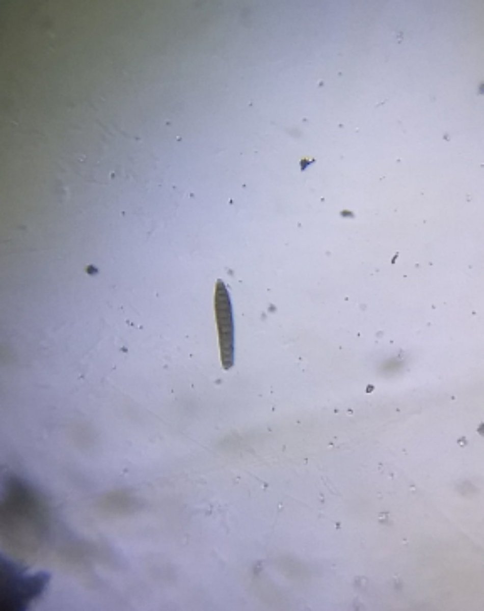
Is this a diatom ?
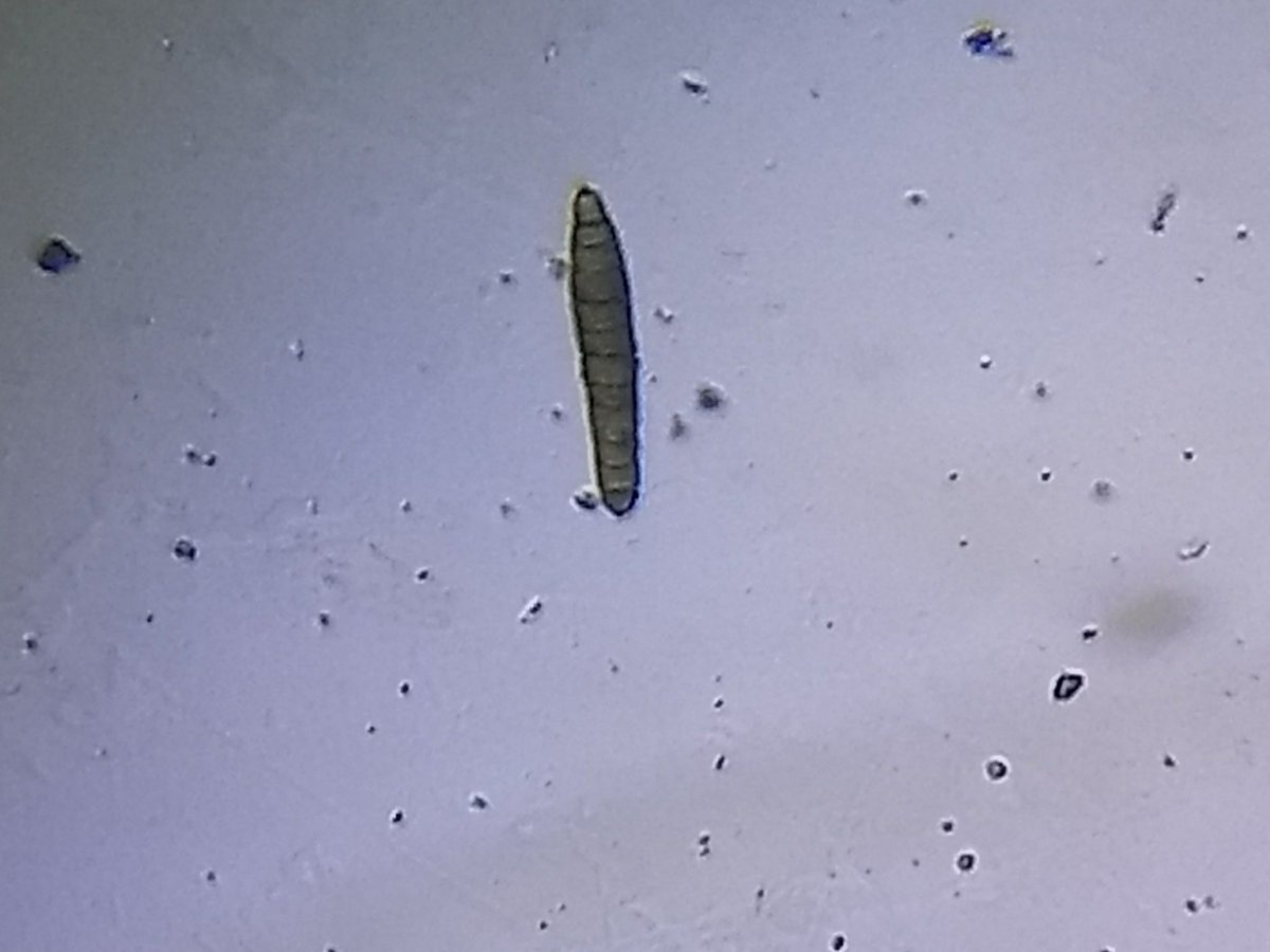
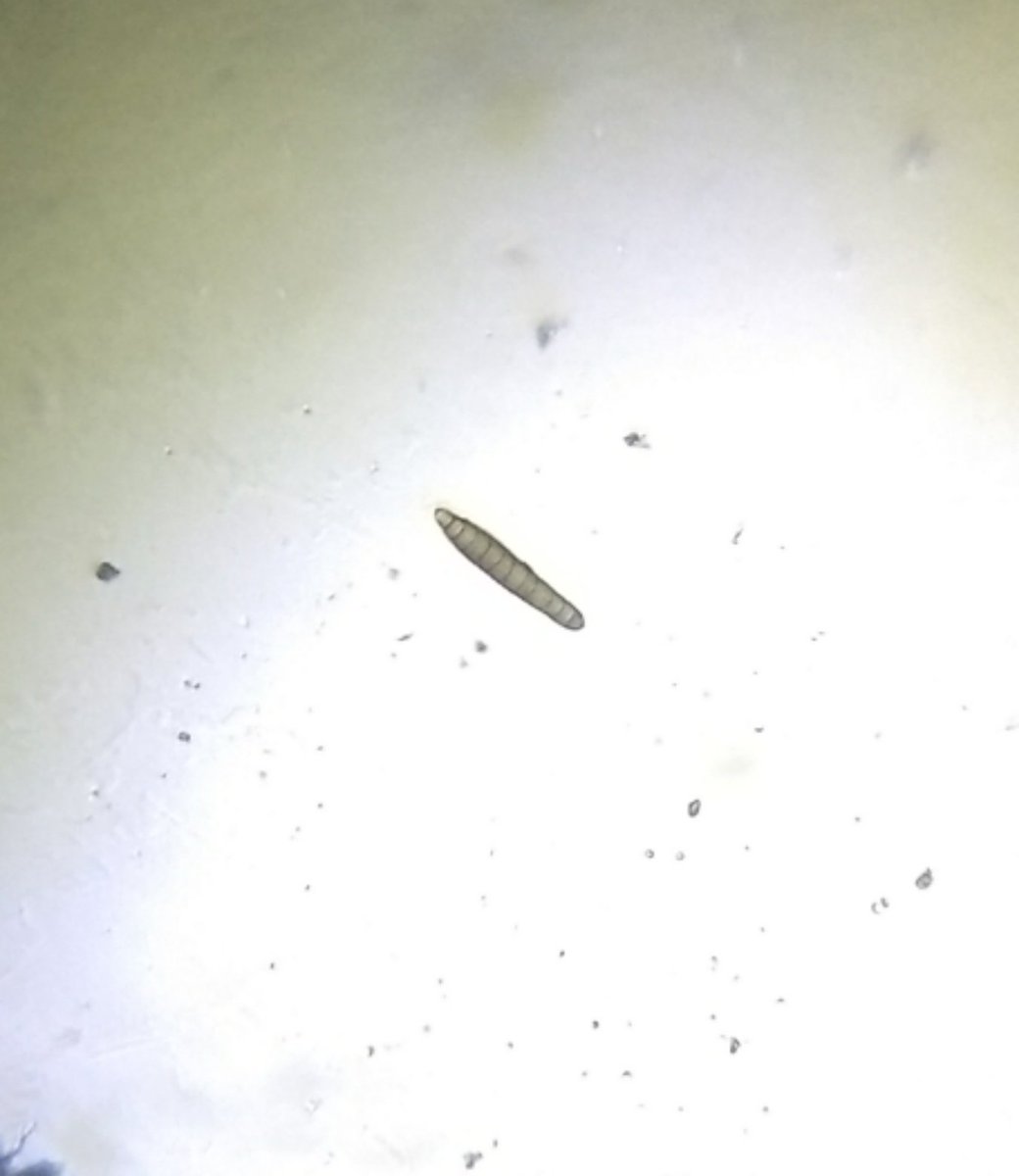
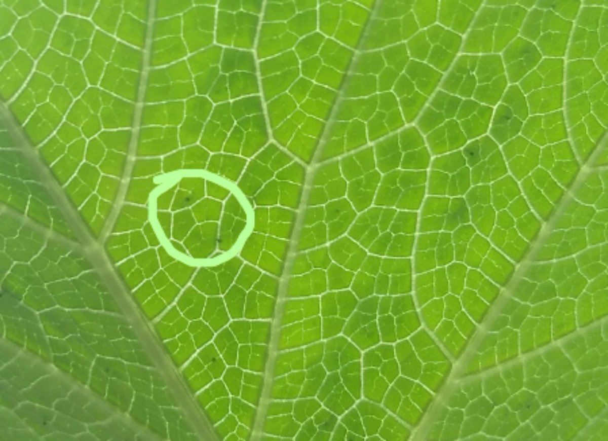
Saw these interesting looking dots on the underside of a pumpkin leaf. Decided to check out using Foldscope. It seemed like it was attached to the leaf and I had to pluck them. Not sure if this is part of plant or something stuck to leaf. Any guesses ?
