Attempted dark field microscopy using a foldscope and a bindi which I borrowed from my wife. Amazed with the results. Checkout the last stomata image. We can almost see 3D structure of the stomata. Feels like electron microscope stuff ..lol
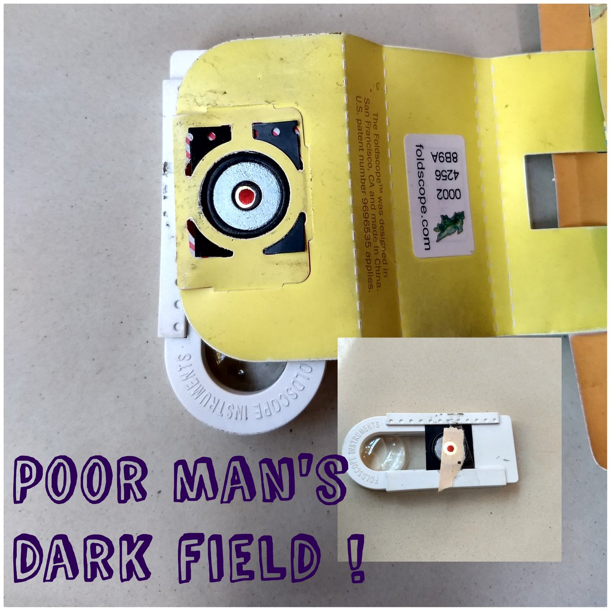 image of 3D structure
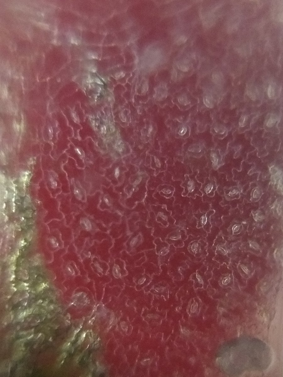 image of 3D structure
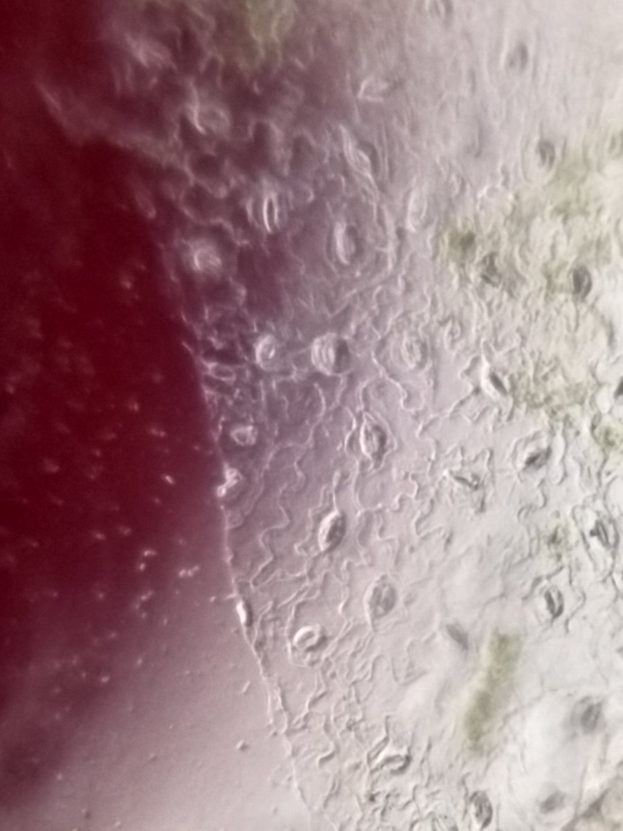 image of 3D structure
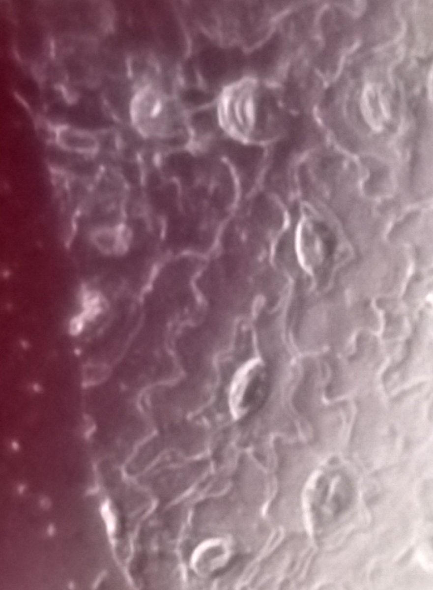 image of 3D structure